StratX® Lung Analysis Platform
Treat with Confidence
A cloud-based quantitative CT analysis service that supports Zephyr® Valve patient selection and treatment targeting by providing information on emphysema destruction, fissure completeness, and lobar volumes
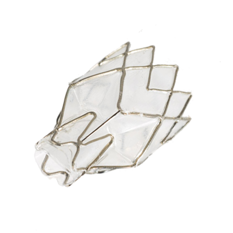
Zephyr Valve treatment is the most rigorously studied MINIMALLY–INVASIVE treatment for severe emphysema and is clinically proven to improve patients’ BREATHING FUNCTION, EXERCISE CAPACITY, and QUALITY OF LIFE.1-3
Lung Report Interpretation
 Summary
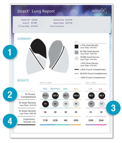
The goal of Zephyr Endobronchial Valve treatment is to completely occlude and reduce the volume of a target lobe, thereby improving hyperinflation and breathing dynamics. In selecting a target lobe, physicians should look for higher levels of emphysema destruction and presence of complete or nearly complete fissures with neighboring lobes (which has been associated with absence of collateral ventilation and likely response to therapy).18
The StratX lung report contains tabulated data on fissure completeness by lobe, destruction score by lobe, and lobar volume. An infographic “key” for easy interpretation of the data is also included. This infographic includes color coding representing the level of emphysema destruction (with darker colors representing lobes with more destruction) and different levels of fissure completeness relative to a target lobe (with darker and more complete lines having greater completeness).
StratX provides fissure completeness, emphysema density, and inspiratory volumes to allow identification of target lobes that are good candidates for treatment with Zephyr Valves.
 Fissure Completeness
Fissure completeness has been shown to be a predictor of success for Zephyr Valve therapy as it indicates absence of collateral ventilation between the target and the neighboring lobes.19
The StratX lung report displays fissure completeness values “by lobe,” i.e., the values are computed as a percentage of the total area of the fissure across the lobar boundary. The fissure completeness between each lobar region is represented with a dark solid, light solid, or dotted line. The dark solid line represents fissures that are ≥95% intact.
The light solid line represents fissures that are 80-95% intact. The dotted line represents fissures which are below 80% intact. A fissure completeness score of <80% indicates the presence of collateral ventilation in that lobe and the lobe should not be considered for treatment with Zephyr Valves. For fissure completeness scores of >80%, fissure integrity should be confirmed using Pulmonx’s Chartis® Pulmonary Assessment System to ensure that the target lobe is negative for collateral ventilation.

Destruction Scoring
Destruction scoring is calculated as the percentage
of voxels in Hounsfield units in a lobe that fall below a threshold value. For example, if the total number of voxels in the lobe was 1000, and 800 voxels fell below the threshold of -910 HU, the “destruction score” for that lobe at -910 HU would be 80%.
Lobar Destruction Score values of greater than 50% at -910 HU have been commonly used as an inclusion criterion for various clinical trials of Zephyr Valve therapy.1-3
In the report and summary view, the degree of shading of a lobe and the numbers within the lobe represent the level of destruction; lobes with less than 50% destruction are colored white and are usually not considered as potential targets. Percent destruction scores below -950 HU are also provided for reference.

Inspiratory Volume
The inspiratory volume represents the volume of each lobe in milliliters. The inspiratory volume can help to identify the lobes with the largest volume representing hyperinflation and ones that may be a good target for Zephyr Valve treatment.
While the summary section provides a good quick reference, the actual values in the results table should be reviewed before making a decision on which lobe(s) to target.
Features a USER-FRIENDLY DESIGN for clear interpretation and ease of use

StratX Features
Lung Analysis Platform Enables You To:
-
Screen treatment candidates non-invasively
-
Choose between multiple potential treatment targets if applicable
-
Enhance case planning and optimize procedure time
-
Compare quantitative analyses across patients
-
Educate referrers about the optimal candidate for Zephyr Valve therapy
-
Educate patients using user-friendly reports
StratX Workflow
Key Scan Parameters
- Ensure all files are in standard .DICOM format. Only supine position chest CT scans are supported. Scans obtained in prone position cannot be analyzed.
- The CT scans must be acquired at a slice thickness of ≤1.5 mm.
- Only TLC (inspiration) scans are required for analysis.
- CT scan should be acquired without contrast in axial view.
- The input image should not be reconstructed with a slice spacing larger than the slice thickness (no gaps in the 3D volume are allowed).
- The complete lungs must be present on the CT scan. If parts of the lung are missing, the output parameters will be compromised.
- Ensure the CT scan is of good quality (e.g., no movement artifacts, no artifacts due to metal, no high noise levels due to dose level, etc.).
- Please ensure the CT scan does not suffer from image artifacts such as streak artifacts from implants.
- Scans taken from CT scanners with less than 16 detector rows are not recommended. Refer to www.pulmonxstratxusa.com or www.pulmonxstratx.com for details regarding scan acquisition parameters.
Complications of the Zephyr Endobronchial Valve treatment can include but are not limited to pneumothorax, worsening of COPD symptoms, hemoptysis, pneumonia, dyspnea and, in rare cases, death.
